
An imaging system helps in tumors detection during surgery
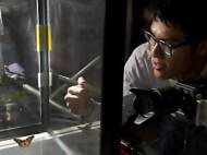
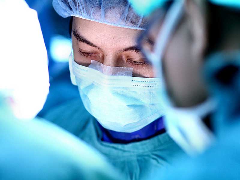 A group of researchers at the Fraunhofer Institute have developed a multispectral fluorescence camera system – a new surgical aid that makes hidden tumors visible during an operation. In near future, this special camera could be integrated into various medical imaging systems, such as surgical microscopes and endoscopes, in order to assist surgeons during tumor removal operations.
Cancer patients have the highest probability of recovery if tumors are completely removed. However, tiny clusters of cancer cells are often difficult for surgeons to recognize and remove. Tumor removal surgeries pose a great challenge even to experienced surgeons because tumor margins are blending into healthy tissue and are difficult to tell apart. Moreover, distributed domains of cancer and pre-malignancies are difficult to recognize.
At present, doctors rely completely on their trained eyes when removing pieces of tumors. The new special camera system developed at Fraunhofer Project Group for Automation in Medicine and Biotechnology (Fraunhofer PAMB) can be used during surgery to help in visualization of even the smallest, easy-to-overlook, malignant pieces of tumor and thereby assist the surgeons during complicated interventions.
Novel camera system can display fluorescent molecules that mark the cancer tissue. These molecules are injected into the patients’ blood circulation before the operation. The molecules selectively attach onto the tumor during their trip through the body. When the corresponding area is illuminated with a specific wavelength, fluorescence is emitted and the malignant tissue glows in the color of the injected dye, while the healthy tissue appears the same. In this way, the surgeon can see clusters of tumors cells that cannot be recognized by the naked eye.

Unlike other imaging systems, novel camera system can display several fluorescent dyes and the reflectance image simultaneously in real time. Arteries and delicate nerves that must not be injured during an intervention can be colored with another dye and detected with the new camera. The detection is possible since they are set apart from their surroundings.
“The visibility of the dye to the camera depends in large part on the selection of the correct set of fluorescence filters. The filter separates the incident excitation wavelengths from the fluorescing wavelengths so that the diseased tissue is also set apart from its surroundings, even at very low light intensities”, said Nikolas Dimitriadis, head of the Biomedical Optics Group at Fraunhofer PAMB.
The research team needed only one camera and one set of filters for their photographs, which can present up to four dyes at the same time. Software is used to analyze and process the images in seconds to present the information continuously on a monitor during surgery. The information from the fluorescent image is overlaid on the normal color image.
“The operator receives significantly more accurate information. Millimeter-sized tumor remnants or metastases that a surgeon would otherwise possibly overlook are recognizable in detail on the monitor. Patients operated under fluorescent light have improved chances of survival”, said Dimitriadis.
According to researchers, their multispectral fluorescence camera can be adapted for use with other available dyes, making it adaptable to various applications in medicine. The system needs to be prepared for such operation.
“One preparation that is already available to make tumors visible is 5-amino levulinic acid (5-ALA). Physicians employ this especially for glioblastomas – one of the most frequent malignant brain tumors in adults. 5-ALA leads to an accumulation of a red dye in the tumor and can likewise be detected with the camera”, said Dimitriadis.
According to the Fraunhofer PAMB researchers, the multispectral fluorescence imaging system should pass testing for use on humans as soon as next year. The first clinical trials with patients suffering from glioblastomas are planned for 2014.


Leave your response!