
Biomimicry of butterfly wings for more powerful solar cells
 The discovery that butterfly wings have scales that act as tiny solar collectors has led scientists in China and Japan to design a more efficient solar cell that could be used for powering homes, businesses, and other applications in the future. The researchers turned to the microscopic solar scales on butterfly wings in their search for improvements. Using natural butterfly wings as a mold or template, they made copies of the solar collectors and transferred those light-harvesting structures to Grätzel cells.Laboratory tests showed that the butterfly wing solar collector absorbed light more efficiently than conventional dye-sensitized cells. The fabrication process is simpler and faster than other methods, and could be used to manufacture other commercially valuable devices, the researchers say
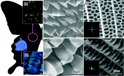
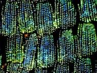
Wings taken from two species of butterfly were used as inspiration for their work, one is Papilio paris Linnaeus (subfamily Papilionidae of the family Nymphalidae), the other is Thaumantis diores (Doubleday) (subfamily Amathusiidae of the family Nymphalidae).
Papilio paris is a species of beautiful swallowtail butterfly found in South China. Expanse of the wings is about 10cm. Upper side of the wings is black, irrorated with dark green scales, which on the outer portion of the forewing coalesce and form an incomplete post discal narrow band. On the hind wing, there is a conspicuous upper discal shining blue patch. The other specie of butterfly used in our work as contrast is Thaumantis diores, the upper wings of which are brown black. All the wings were cut into 1cm × 1cm square samples from different colors.
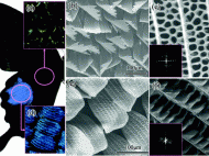
A four-level observation method was adopted in studying the morphology of butterfly wings. The first level deals with the macroscopic aspect of the butterfly wings. The second level is the optical microscopy observation level. The outline and color of the scales can be clearly identified in these images. FESEM was used to study the fine structures (the third and fourth level) of the wing scales. For FESEM measurement, all the samples were stuck to microscope stubs with double-sided carbon tape and then coated by a thin, sputtered gold layer to provide a conducting surface and avoid charging effects. It should be noted that previous studies have proven that there is no significant distortion of the scale geometry during this process.

An electrically conducting Fluorine-doped Tin Oxide (FTO) coated glass was used as substrate. The FTO glass was cleaned in deionized water with ultrasonication for 5 minutes. It was then hydroxylated by ultrasonication for 3 minutes in a mixture of 60ml of isopropanol and 40ml of aqueous KOH with concentration of 1M, followed by rinsing in deionized water with sonication for 10 min. After these cleaning procedures, the surface of substrates became hydrophilic. It can be anticipated that the hydrophilic FTO glass is suitable for covering a layer of TiO2 film.
Titanium sulfate was dissolved in absolute alcohol with different concentrations. After the solution was stirred for 1h at 60°C, the pH was adjusted to the range of 2.5−3.0 by adding dilute H2SO4. An appropriate amount of nonionic surfactant Triton X100 was also added to this solution. Butterfly wings were pretreated in advance to remove salts and proteins. Then the pretreated butterfly wings were immersed into the titanium sulfate precursor solution maintained at constant temperature over 60°C for 24h or more, and then removed from the precursor solution, rinsed thoroughly with absolute alcohol. At first, we prepared a layer of colloidal TiO2 dispersed on the FTO conductive surface. The immerged wing scales were then subsequently placed on the layer of the colloidal TiO2 film, covered by another piece of FTO glass. Two pieces of the FTO glasses were tightly clamped, as to keep the soaked wings flat and improve the interface between colloidal TiO2 film layer and soaked biotemplates.

At last, the specimen was then placed into an oven, and was heated up to 500°C with the slow heating rate at 1°C/min in air. The slow heating rate was adopted so that to avoid the appearance of cracks and frame collapse of the soaked wings during sintering process. By keeping the specimen inside for 2h with such temperature, the chitinous substrates were removed completely after reaction with air, leaving only TiO2 in the form of ceramic butterfly wings. Then, the as-prepared “butterfly wing microstructure photoanode” was thus obtained. The as-synthesized photoanodes are divided into four layers: glass substrates, F:SnO2 conductive layer, anatase film, and titania film with butterfly wing microstructures.
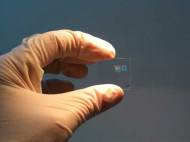
In the study, Di Zhang and colleagues note that scientists are searching for new materials to improve light-harvesting in so-called dye-sensitized solar cells, also known as Grätzel cells for inventor Michael Grätzel. These cells have the highest light-conversion efficiencies among all solar cells (as high as 10 percent).


What a great adoption of marvels of nature to improve the efficiency of solar cells.
Dr.A.Jagadeesh Nellore(AP),India
Nice article, still lot to learn from biological nano-structures. It would be great if nanostuctures could easily implemented in large scale.. .
Blenson Paul.
TU-Dresden, Germany.
Mangalore, India.