
Improved printing of cartilage replacement constructs
 The printing of 3D tissue has taken a major step forward with the creation of a novel hybrid printer that simplifies the process of creating implantable cartilage. Researchers from the Wake Forest Institute for Regenerative Medicine were able to use this technology in order to create cartilage constructs that could eventually be used to replace or to induce re-growth of cartilage in specific areas of injured patients.
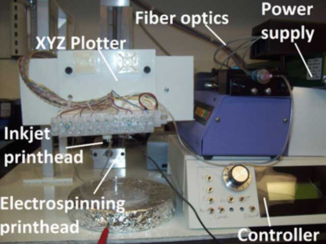
The printer is a combination of two low-cost fabrication techniques – a traditional ink jet printer and an electrospinning machine. Combination of these systems allowed the researchers to build a structure made from natural and synthetic materials. Synthetic materials ensure the strength of the construct and natural gel materials provide an environment that promotes cell growth.
In comparison to cartilage created by an ink jet printer using gel material alone, the hybrid system produces cartilage constructs with increased mechanical stability in both lab and real-life systems. This success was achieved due to use of the electrospinning machine, which employs electrical current to generate very fine fibers from a polymer solution. Electrospinning allows the composition of polymers to be easily controlled and thus produces porous structures that encourage cells to integrate into surrounding tissue.
“This is a proof of concept study and illustrates that a combination of materials and fabrication methods generates durable implantable constructs”, said James Yoo, M.D., Ph.D., Professor at the Wake Forest Institute for Regenerative Medicine. “Other methods of fabrication, such as robotic systems, are currently being developed to further improve the production of implantable tissue constructs.”
In order to prove their concept, the researchers applied alternate layers of flexible mats of electrospun synthetic polymer and a solution of cartilage cells from a rabbit ear that were deposited using the traditional ink jet printer.

The resulting constructs have 100 × 100 × 0.4 mm (3.94 × 3.94 × 0.015 inches), and the researchers tested their strength by loading them with variable weights and tested whether the cartilage cells are alive after one week. The constructs were also implanted into mice for 2, 4 and 8 weeks to see how they performed in a real life system. The results after 8 weeks of implantation have shown that the constructs have developed the structures and properties of elastic cartilage.
Wake Forest Institute for Regenerative Medicine researchers envision their technology to be used along with data gathered by MRI scans. After the desired part of the body with the need for cartilage repairment is scanned by MRI, the data could be used as a blueprint for creation of a matching cartilage construct.
A careful selection of scaffold material for each patient’s construct would allow the implant to withstand mechanical forces while encouraging new cartilage to re-grow at the desired area. Further refinement of the technique could produce functional cartilage constructs by using oriented fibers to direct chondrocyte growth.
For more information, you can read the paper published in the IOP Publishing’s journal Biofabrication: “Hybrid printing of mechanically and biologically improved constructs for cartilage tissue engineering applications”.
Share:

-
-
-
-
-
-


Leave your response!