
Moth-eyes biomimicry may enhance X-ray imaging
 Using the compound eyes of the humble moth as their inspiration, an international team of physicists from the City University of New York and Tongji University in Shanghai applied biomimicry to develop new nanoscale materials that could someday increase the resolution of the resulting X-ray images without the need for larger radiation dosages which occur due to amplification of input radiation.
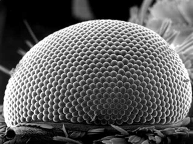
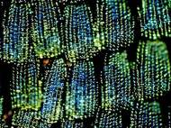
Moths have large compound eyes which consist out of many thousands of ommatidia-structures which form a primitive cornea and lens that are connected to photoreceptor cells. Their eyes are also anti-reflective and bounce back very little of the light that strikes them in order to help the insects be stealthier and less visible to predators during their nocturnal flights. Because of this feature, engineers have looked to the moth eye to help design more efficient coatings for solar panels and displays.
Led by Yasha Yi, a professor of the City University of New York, who is also affiliated with MIT and New York University, the researchers took another path and used the moth eye as a model for a new class of materials that improve the light-capturing efficiency of X-ray machines and similar medical imaging devices.

In particular, the researchers focused on so-called “scintillation” materials: compounds that, when struck by incoming particles (say, X-ray photons), absorb the energy of the particles and then reemit that absorbed energy in the form of light. In radiographic imaging devices, such scintillators are used to convert the X-rays exiting the body into the visible light signals picked up by a detector to form an image.
When you increase the amount of radiation the resulting image resolution gets better, but that approach isn’t healthy for patients. On the other hand, you could increase the sensitivity of the sensor in order to achieve better resulting image and Yi and colleagues figured that could be achieved with improvement of the scintillator – a part of the X-ray machine which converts X-rays to light.
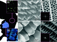
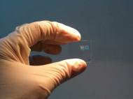
The new material developed for the scintillator consists of a 500 nanometers thick film made of a special type of crystal known as cerium-doped lutetium oxyorthosilicate. These crystals were encrusted with tiny silicon nitride pyramid-shaped bumps which are modeled after the structures in a moth’s eye in order to extract more light from the film.
The achieved density of the bumps is about the same as in an actual moth eye – between 100,000 to 200,000 fit within a 100 x 100 micrometer square. The researchers then made the sidewalls of the device rougher, improving its ability to scatter light and thus enhanced the efficiency of the scintillator.
In lab experiments, Yi and colleagues found that adding the thin film to the scintillator of an X-ray mammographic unit increased the intensity of the emitted light by as much as 175 percent compared to that produced using a traditional scintillator.
The current work, Yi says, represents a proof-of-concept evaluation of the use of the moth-eye-based nanostructures in medical imaging materials.
“The moth eye has been considered one of the most exciting bio structures because of its unique nano-optical properties”, said Yi, “and our work further improved upon this fascinating structure and demonstrated its use in medical imaging materials, where it promises to achieve lower patient radiation doses, higher-resolution imaging of human organs, and even smaller-scale medical imaging. And because the film is on the scintillator the patient would not be aware of it at all.”
The researches are going to work with medical imaging experts and radiologists to make this technology implemented into actual clinical practice, and Yi estimates that it will take at least another three to five years to evaluate and perfect the film.
For more information, read the paper published in the Optical Society’s (OSA) journal Optics Letters: “Giant light extraction enhancement of medical imaging scintillation materials using biologically inspired integrated nanostructures”.
Share:

-
-
-
-
-
-


Leave your response!