
Single GFP-expressing cell is basis of a living biological laser device
 Two researchers at the Wellman Center for Photomedicine at Massachusetts General Hospital have developed a living laser, in which a single cell genetically engineered to express green fluorescent protein (GFP). Before you start thinking about sharks with lasers or other science fiction creatures, this technology is used to amplify photons into nanosecond-long pulses of laser light.
“Since they were first developed some 50 years ago, lasers have used synthetic materials such as crystals, dyes and purified gases as optical gain media, within which photon pulses are amplified as they bounces back and forth between two mirrors”, said Seok Hyun Yun, an associate professor of Dermatology at Harvard Medical School. “Ours is the first report of a successful biological laser based on a single, living cell.”
The researchers chose GFP (a protein originally found in a species of jellyfish) because it can be induced to emit light without the application of additional enzymes. Its properties are well understood, and there are established techniques to genetically program many organisms to express GFP. To determine the protein’s potential for generating laser light, the researchers first assembled a device consisting of a 2.5cm (1 inch) long cylinder, with mirrors at each end, filled with a solution of GFP in water. After confirming that the GFP solution could amplify input energy into brief pulses of laser light, the researchers estimated the concentration of GFP required to produce the laser effect.

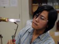
Their next step was to develop a line of mammalian cells expressing GFP at the required levels. The cellular laser was assembled by placing a single GFP-expressing cell (with a diameter ranging from 15 to 20 micrometers) in a microcavity consisting of two highly reflective mirrors spaced 20 micrometers apart. Aside being able to produce pulses of laser light in the GFP solution experiment, the researchers found that the spherical shape of the cell itself acted as a lens which refocuses the light and induces emission of laser light at lower energy levels than required for the solution-based device. The cells used in the device survived the lasing process and were able to continue producing hundreds of pulses of laser light.
“One of our long-term goals will be finding ways to bring optical communications and computing, currently done with inanimate electronic devices, into the realm of biotechnology. That could be particularly useful in projects requiring the interfacing of electronics with biological organisms. We also hope to be able to implant a structure equivalent to the mirrored chamber right into a cell, which would the next milestone in this research”, said Malte Gather, Research Fellow in Dermatology at Massachusetts General Hospital.
Although the individual laser pulses last for only a few nanoseconds, they are bright enough to be readily detected and carry information that could help in examination of large numbers of cells. This technology could lead to new forms of body imaging, and the laser could be used in photodynamic therapies, where a biocompatible source placed inside a patient could be used to activate the drugs.
For more information, you can read the article published in the journal Nature Photonics named: “Single-cell biological lasers”.


i enjoy when you add a tone of humor inside these scientific articles. It lights up the rest of the read.