
Imaging nanoscale dynamic systems in structural biology
 Seeing viruses in action in their natural environment is invaluable, and researchers at the Virginia Tech Carilion Research Institute have devised a way to directly image biological structures at their most fundamental level and in their natural habitats. The technique is a major advancement toward the ultimate goal to image nanoscale dynamic systems in structural biology while being in action.
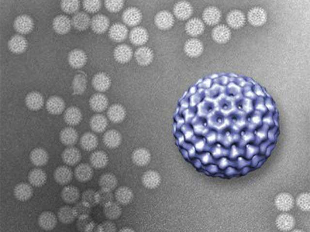
The researchers used rotavirus to test their imaging technique. Rotavirus is the most common cause of severe diarrhea among infants and children, and although the disease tends to be easily managed in the developed world, in developing countries rotavirus kills more than 450,000 children a year.
At the second step in the life cycle of rotavirus, the virus sheds its outer layer and becomes what is called a double-layered particle. Smaller size of the double-layered particle allows it to enter a cell and begin using the cell’s own infrastructure to produce more viruses. Sarah McDonald, an assistant professor at the Virginia Tech Carilion Research Institute, provided a pure sample of rotavirus double-layered particles for the study.
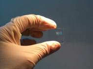
The technique involves taking two silicon-nitride microchips with windows etched in their centers and pressing them together until they are only 150 nanometers apart. This space is filled with a liquid which serves as a microfluidic chamber which functions as the natural environment of the observed biological structure. Microchip’s interior surface is coated with a layer of natural biological tethers, such as antibodies, which naturally grab onto a virus and hold it in place in order to gain better quality of images.
Led by Deborah Kelly, an assistant professor at the Virginia Tech Carilion Research Institute, the researchers coated the interior window of the microchip with antibodies which were utilized to grab the rotaviruses that were injected into the microfluidic chamber and hold them in place. After preparation, the researchers used a transmission electron microscope to image the slide, and the experiment revealed results similar to those achieved by traditional freezing methods used to prepare rotavirus for electron microscopy.
After creating a method to record deliver accurate results in non-frozen conditions, Virginia Tech researchers plan to improve the technique with a goal to observe biological structures dynamically in action. Future results could help McDonald and her colleagues to understand how rotavirus assembles, thus enabling development of means to fight this virus.
For more information, you can read the article published in the Lab on a Chip: “Visualizing viral assemblies in a nanoscale biosphere” [605KB PDF].


Leave your response!