
Researchers use nanotechnology to create a nanopatch for the heart
 After a heart attack, the nerve cells in the heart’s wall and cells that keep the heart beating in perfect synchronicity are lost forever. The best approach would be to figure out how to revive the deadened area, and a group of researchers at the Brown University and the India Institute of Technology Kanpur turned to nanotechnology in order to find a solution in a form of a nanopatch.
In 2009, some 785,000 Americans suffered a new heart attack linked to weakness caused by the scarred cardiac muscle from a previous heart attack, according to the American Heart Association. According to American Heart Association, a third of women and a fifth of men who have experienced a heart attack will have another one within six years.
“This whole idea is to put something where dead tissue is to help regenerate it, so that you eventually have a healthy heart”, said David Stout, a graduate student in the School of Engineering at Brown.
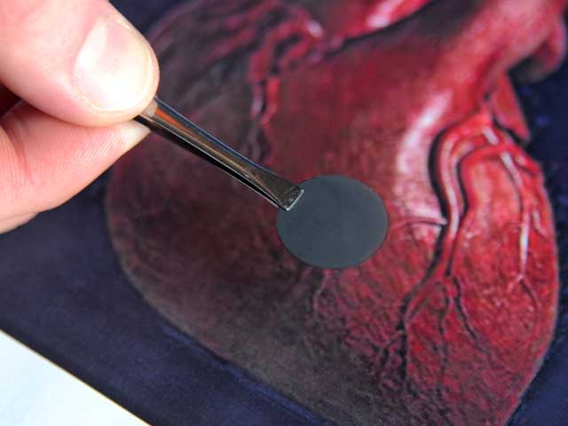
In a lab, the researchers built a scaffold-looking structure consisting of carbon nanofibers and a government-approved polymer. Tests showed the synthetic nanopatch regenerated natural heart tissue cells (cardiomyocytes) as well as neurons, thus reviving a dead region of the heart. The natural heart-tissue cell density on the nanoscaffold was six times greater than the control sample, while neuron density had doubled.
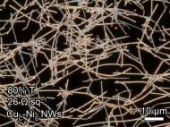
What is unique about the experiments at Brown University and at the India Institute of Technology Kanpur is the use of carbon nanofibers, helical-shaped tubes with diameters between 60 and 200 nanometers. The carbon nanofibers work well because they are excellent conductors of electrons, performing the kind of electrical connections the heart relies upon for keeping a steady beat.
The researchers stitched the nanofibers together using a poly lactic-co-glycolic acid (PLGA) polymer to form a mesh about 22 millimeters long and 15 microns thick and resembling a patch. They laid the mesh on a glass substrate to test whether cardiomyocytes would colonize the surface and grow more cells.
In tests with the 200-nanometer-diameter carbon nanofibers seeded with cardiomyocytes, five times as many heart-tissue cells colonized the surface after four hours than with a control sample consisting of the polymer only. After five days, the density of the surface was six times greater than the control sample, the researchers reported. Neuron density had also doubled after four days, they added.
“The scaffold works because it is elastic and durable, and can thus expand and contract much like heart tissue”, said Thomas Webster, associate professor in engineering and orthopaedics at Brown. “It’s because of these properties and the carbon nanofibers that cardiomyocytes and neurons congregate on the scaffold and spawn new cells, in effect regenerating the area.”
The scientists want to tweak the scaffold pattern to better mimic the electrical current of the heart, as well as build an in-vitro model to test how the material reacts to the heart’s voltage and beat regime. They also want to make sure the cardiomyocytes that grow on the scaffolds are endowed with the same abilities as other heart-tissue cells.
For more information, you can read the article published in the Acta Biomaterialia named: “Poly(lactic–co-glycolic acid): Carbon nanofiber composites for myocardial tissue engineering applications”.


Leave your response!