
Biomedical breakthrough – blood vessels for lab-grown tissues
 Researchers from Rice University and Baylor College of Medicine (BCM) have broken one of the major roadblocks on the path to growing transplantable tissue in the lab, because they found a way to grow the blood vessels and capillaries needed to keep tissues alive. In a combination with their previous research, the team could grow blood vessels in predetermined patterns.
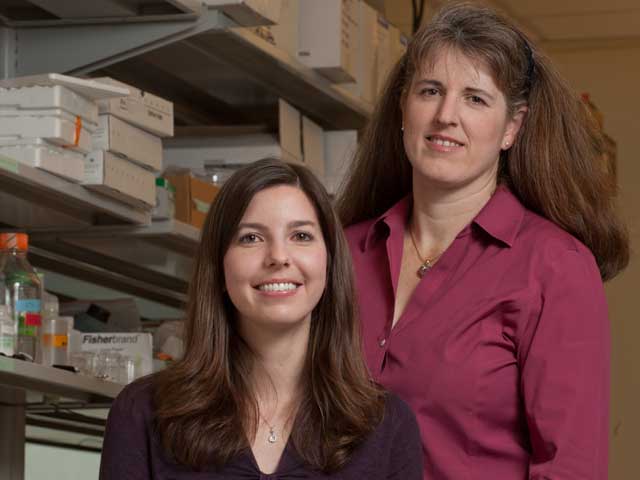
“The inability to grow blood-vessel networks – or vasculature – in lab-grown tissues is the leading problem in regenerative medicine today”, said Jennifer West, department chair and the Isabel C. Cameron Professor of Bioengineering at Rice. “If you don’t have blood supply, you cannot make a tissue structure that is thicker than a couple hundred microns.”
As its base material, a team of researchers led by West and BCM molecular physiologist Mary Dickinson chose polyethylene glycol (PEG), a nontoxic plastic that’s widely used in medical devices and food. Building on 10 years of research in West’s lab, the scientists modified the PEG to mimic the body’s extracellular matrix (network of proteins and polysaccharides that make up a substantial portion of most tissues).
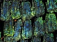
In order to achieve the blood-vessel formation, the researchers combined the modified PEG with two kinds of cells. By using light that locks the PEG polymer strands into a solid gel, they created soft hydrogels that contained living cells and growth factors. After that, they filmed the hydrogels for 72 hours. By tagging each type of cell with a different colored fluorescent marker, the team was able to watch as the cells gradually formed capillaries throughout the soft, plastic gel.
To test these new vascular networks, the team implanted the hydrogels into the corneas of mice, where no natural vasculature exists. After injecting a dye into the mice’s bloodstream, the researchers confirmed normal blood flow in the newly grown capillaries.
Another key advance, published by West and graduate student Joseph Hoffmann in November, involved the creation of a new technique called “two-photon lithography” – a delicate way of using light to create complex 3D patterns within the soft PEG hydrogels.
West said the patterning technique allows the engineers to use a fine level of control over where cells move and grow. In their future experiments, West and her team plan to use the technique to grow blood vessels in predetermined patterns. More information can be found in a paper published in the January issue of journal Acta Biomaterialia named: “Covalently immobilized platelet-derived growth factor-BB promotes angiogenesis in biomimetic poly(ethylene glycol) hydrogels”.


This is great news, congratulations too the reseraches associated with the project! We are now one step closer to true lab grown tissue.