
3D printed vascular network templates made from sugar
 Researchers from the University of Pennsylvania have developed an innovative solution that allows creation of 3D printed templates of filament networks which can be used to rapidly create vasculature and improve the function of engineered living tissues. Unlike other approaches which involve 3D printing in layers, Penn researchers decided to accelerate this delicate process and turned the printing process inside out.
The research was conducted by a team led by postdoctoral fellow Jordan S. Miller and Christopher S. Chen, the Skirkanich Professor of Innovation in the Department of Bioengineering at Penn, along with Sangeeta N. Bhatia, Wilson Professor at the MIT, and postdoctoral fellow Kelly R. Stevens in Bhatia’s laboratory.
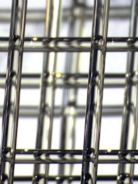
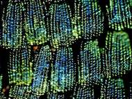
Instead using the layer-by-layer approach, Chen and colleagues focused on the vasculature layout and designed free-standing 3D filament networks in the shape of a vascular system that sat inside a mold. The approach allows removal of the mold and vascular template once the added cells formed a solid tissue enveloping the filaments.
“Sometimes the simplest solutions come from going back to basics”, said Miller. “I got the first hint at this solution when I visited a Body Worlds exhibit, where you can see plastic casts of free-standing, whole organ vasculature.”
This new rapid casting technique is possible due to the development of a material that is suitable for use in 3D printers, with properties which allow it to be rigid enough to form a 3D network of cylindrical filaments which can also be easily removed without toxic effect once the cells form solid tissue around it.
After much testing, the team found the perfect mix of material properties in a humble material – sugar. While sugars crystals are mechanically strong and make up the majority of organic biomass on the planet in the form of cellulose, their building blocks are also typically added and dissolved into nutrient media that help cells grow.
“We tested many different sugar formulations until we were able to optimize all of these characteristics together”, said Miller. “Since there’s no single type of gel that’s going to be optimal for every kind of engineered tissue, we also wanted to develop a sugar formula that would be broadly compatible with any cell type or water-based gel.”
The formula they settled on is a combination of sucrose and glucose along with dextran for structural reinforcement. An important step in stabilizing the sugar after 3D printing is to coat the templates in a thin layer of a degradable polymer derived from corn. This coating allows the sugar template to be dissolved and to flow out of the gel through the channels they create without inhibiting the solidification of the gel or damaging the growing cells nearby.
Once the sugar is removed, the researchers start flowing fluid through the vascular architecture and cells are able to receive nutrients and oxygen just like it naturally happens in the body. The experiment showed that human blood vessel cells injected throughout the vascular networks spontaneously generated new capillary sprouts to increase the network’s reach, much in the way blood vessels in the body naturally grow.
Another advantage of this system is its ability to create gels containing primary liver cells to test whether their technique could improve their function. When the researchers pumped nutrient-rich media through the gel’s template-fashioned vascular system, the entrapped liver cells boosted their production of albumin and urea (natural components of blood and urine, respectively) which are important measures of liver-cell function and health. There was also clear evidence of increased cell survival around the perfused vascular channels.
Though these engineered tissues were not equivalent to a fully functioning liver, the researchers used cell densities that approached clinical relevance, suggesting that their printed vascular system could eventually be used to further research in lab-grown organs and organoids.
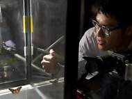
3D printing was done on RepRap, an open-source 3D printer with a custom-designed extruder and controlling software. The whole process is quick and inexpensive, allowing the researchers to switch with ease between computer simulations and physical models of multiple vascular configurations. Several of the custom parts of the RepRap printer the researchers used to make the vascular templates were printed in plastic on another RepRap.
“We want to redesign the printer from scratch and focus it entirely on cell biology, tissue engineering and regenerative medicine applications”, Miller said.
For more information, read the paper published in the journal Nature Materials: “Rapid casting of patterned vascular networks for perfusable engineered three-dimensional tissues”.


Leave your response!